
Precision and permanence: Engineering living neural interfaces
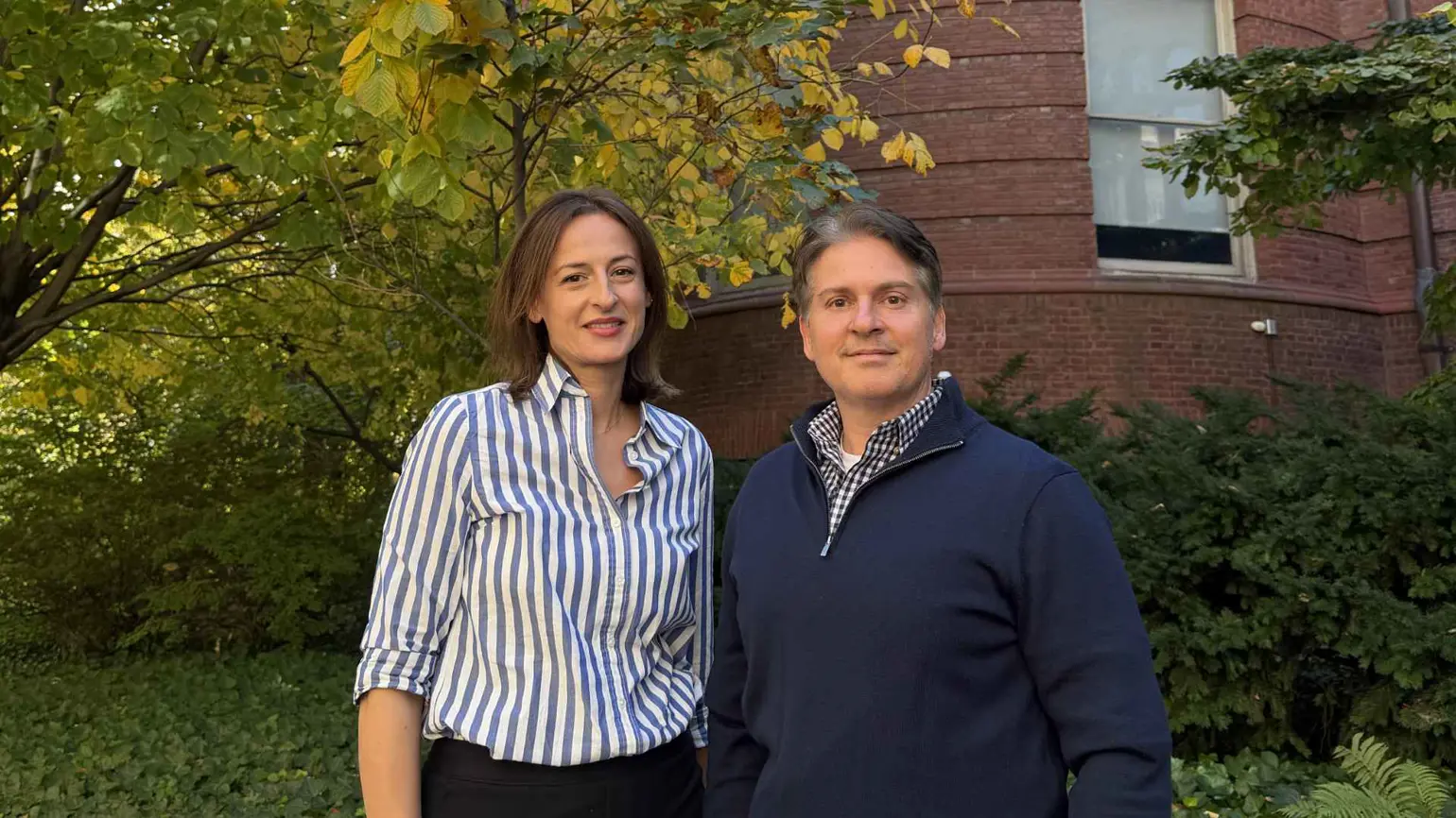
Breakthroughs rarely come from a single insight or a single lab. Professor Kacy Cullen’s team are working on new kinds of biohybrid devices as part of our Precision Neurotechnologies programme. Building a living neural interface involves tissue engineering, biomaterials, optogenetics, animal research, neurosurgery and clinical practice — each tackling a different part of the same problem. Together, the team is asking whether they can create a controllable, long-lasting connection to the brain that could one day offer a different approach to treating Parkinson’s disease.
Deep brain stimulation (DBS) has been used to treat advanced Parkinson’s disease for thirty years. The device sends controlled, mild electrical pulses to specific parts of the brain, helping to control movement symptoms when medication is no longer effective. It’s also a recognised treatment for epilepsy and is increasingly being trialled for treatment-resistant depression. But DBS has its limitations, as Professor Kacy Cullen puts it, “an electrical shock is not really the language that the brain speaks”. If a living tissue can be engineered to successfully connect to the brain’s circuitry, it could engage with the brain’s native chemistry and function as part of that circuit over the long term, without the problems of scarring or device failure.
“We asked the question: is it possible to have something like deep brain stimulation, but build it out of living neurological tissue?”
In Parkinson’s disease, the neurons that produce dopamine degenerate, interfering with the circuits of the brain that regulate movement. Professor Cullen’s team is developing a novel approach. Rather than stimulating these brain regions with electricity, they’re growing dopaminergic neurons that have long axonal fibres – these fibres help neurons communicate across and between brain regions. The aim is that these engineered cells form synapses – the junctions between neurons – and ultimately deliver dopamine signals into the target circuit.
These cells need to be very precisely engineered. This project is managed by Dr Dimple Chouhan, a biomanufacturing expert. She grows these tissues in tiny micro-columns, with a cluster of neurons at one end with a long trail of axons extending from it. Her team have already grown versions of these cells, with axonal trails of two to three centimetres – long enough to bridge the gap between critical brain regions affected in Parkinson’s. The next challenge for the team is showing that this approach works in large-animal studies.
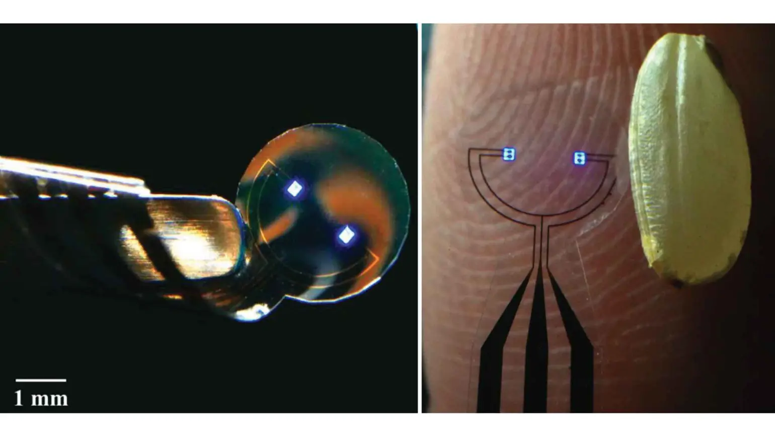
Two blue micro-LEDs on a flexible electrode array, shown alongside a grain of rice for scale.
Designing a controllable system
For these living implants to be effective, they need to be controllable. This is where Professor Flavia Vitale comes in. Her team are using optogenetics to modulate these cells by connecting them to miniature microLED arrays that sit on the surface of the brain. The cells are modulated using specific light wavelengths that either activate or inhibit their activity. These engineered neurons are made light-responsive during the manufacturing process, meaning they don’t influence the surrounding cells in the patient’s brain. The team’s wider ambition is a system that can both read and write brain activity by monitoring and modulating the living implant.
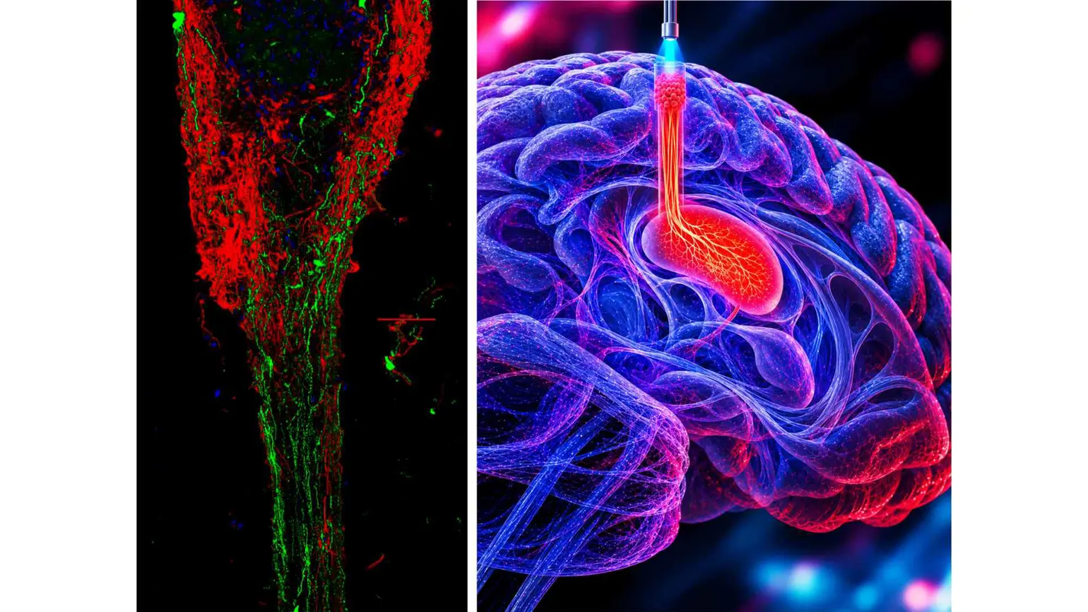
Left: Fluorescent microscopy image of a tissue-engineered living electrode, highlighting its cellular structure. Right hand size: Illustration of a tissue-engineered living electrode integrated within brain tissue.
Because the tissue is grown outside the body, it can be tested for its structure, health and response to light before implantation. “Think of a manufacturing plant building airplanes,” Cullen says. “Before that first flight, there is a lot of quality control. We can do similar quality control for the structural health and light-based functionality of these cells before they are implanted.”
A therapy built by many disciplines
Moving this bio-hybrid device from the lab and into larger mammals and ultimately humans means it needs to be designed with translation in mind. Central to this is the animal-research programme led by Dr Kevin Browne. He works closely with two neurosurgeons, Dr Mykhailo Tatarchuk and Dr Isaac Chen, who ensure the team learns from each study the lessons it will need to ultimately deliver this technology to humans. Dr John Duda, a neurologist and movement-disorder specialist, brings the perspective of the patients and medical conditions the team hopes ultimately to treat.
Together, the team is trying to solve every part of the problem at once: how to manufacture a living neural connection, keep it healthy, control it precisely, implant it safely and prove that it restores meaningful function.
Learning together at the frontier
Cullen’s project is part of a wider group of ARIA-funded teams exploring biohybrid neurotechnologies: systems that combine living biological components with advanced devices.
Within the programme, the teams share protocols, compare approaches and common challenges. “It really feels like a partnership,” Cullen says of the ARIA model. “We all have a great place that we want to get to. Let’s all help each other get there.”
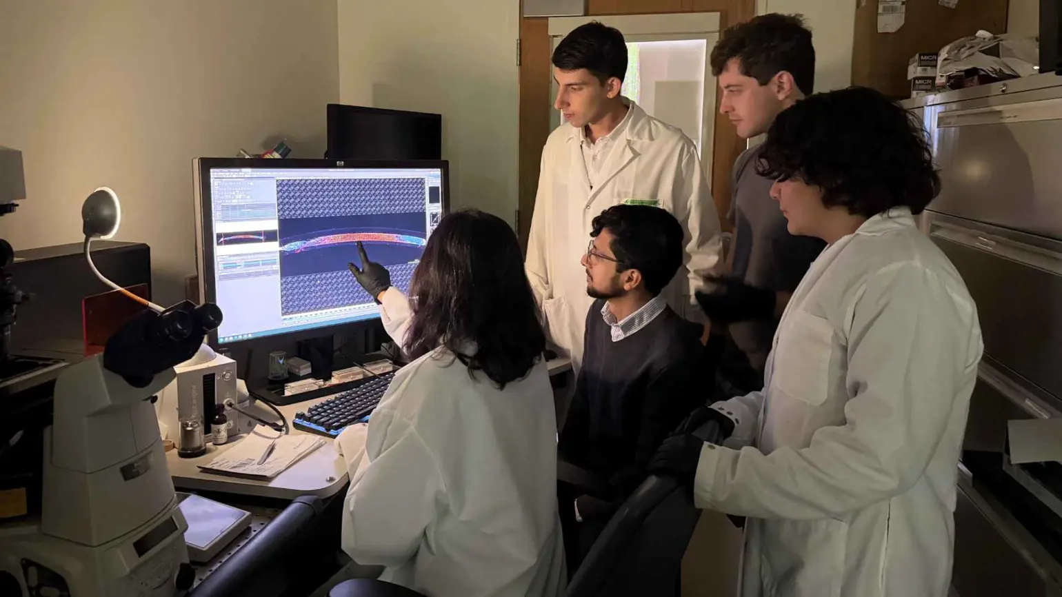
The team in the microscope room.
The work ahead is difficult. It requires new ways to manufacture living tissue, validate its performance and show that it can operate safely over time. But if it succeeds, this team will have achieved a significant shift in what a neural interface can be: from a device that simply delivers a signal to the brain to a living connection capable of reconnecting the brain’s natural pathways and chemistry.